Home
/ Labeled Plant Cell Under Electron Microscope : Ultrastructure - The cell is the basic structural and functional unit of life in all living organisms.
Labeled Plant Cell Under Electron Microscope : Ultrastructure - The cell is the basic structural and functional unit of life in all living organisms.
Labeled Plant Cell Under Electron Microscope : Ultrastructure - The cell is the basic structural and functional unit of life in all living organisms.. Now the first thing to point out when looking at images under an electron microscope is the scale. Plant cells have cell walls, one large vacuole per cell, and chloroplasts, while animal cells will have a cell membrane only. Microscope images in this course come from the light microscope (magnification up to 400x) and the electron microscope (magnification up to 500000x). Learn the structure of animal cell and plant cell under light microscope. Some organelles are visible with a compound light microscope, while other organelles can be seen only under a more powerful tool, such as an electron.
Labeled animal cell under electron microscope. The microscope has been a powerful tool for studying cellular details and phenomena for hundreds of years ever since the first cells, cork cells of plants, were observed by robert hooke in the 1600s. Only light from the plane of focus reaches the detector. Cell biology (biomedical laboratory science students). Diagram of plant cell under microscope.
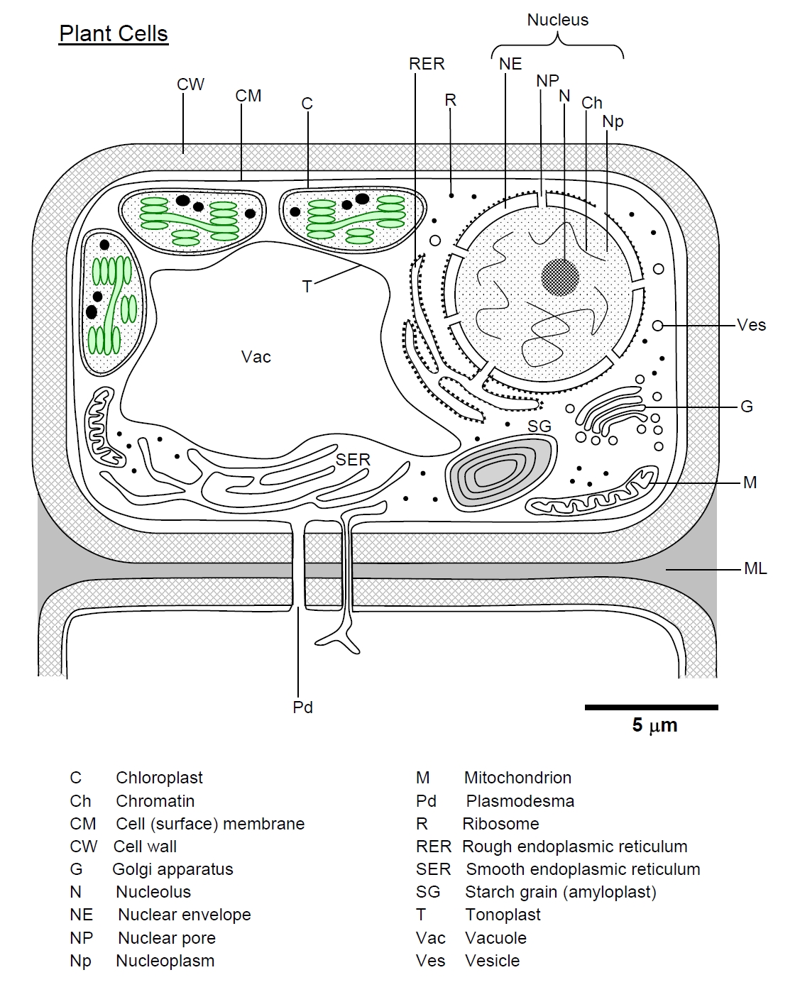
Plant Bodies Cells from cronodon.com Examining plant cells under the microscope. Typical animal cell pinocytotic vesicle lysosome golgi vesicles golgi vesicles rough er (endoplasmic reticulum) smooth er (no ribosomes) cell (plasma) membrane mitochondrion golgi apparatus nucleolus nucleus centrioles (2) each composed of 9 microtubule triplets microtubules cytoplasm ribosome The cell membrane, also known as plasma membrane or plasmalemma consists of three layers when viewed under the electron microscope. Learn the structure of animal cell and plant cell under light microscope. Both plant and animal cells are surrounded by a cell membrane composed of lipids and proteins. Are plant cells prokaryotic or eukaryotic? All information about labeled plant cell under electron microscope. The cell organelles are seen as tiny dots throughout the cytoplasm, whereas the nucleus is seen as a thick drop.
Diagram of plant cell under microscope.
Electron microscopes use a beam of electrons instead of beams or rays of light. The cell membrane, also known as plasma membrane or plasmalemma consists of three layers when viewed under the electron microscope. Using a microscope, it's possible toview and identify these cells and how they are arranged (epidermal cells,spongy cells etc). Here's a photo of a plant cell under an electron microscope. For many years, until the electron microscope was invented, this was the limit of how much we could know about the cell. Cell structure teaching resources the science teacher, organelles biology for majors i, 11 different types of cells in the human body, class test, chronic inflammation under the microscope learn share. Draw and label an animal cell as seen under an electron microscope. Plant cell labeled under microscope : The cell organelles are seen as tiny dots throughout the cytoplasm, whereas the nucleus is seen as a thick drop. (1) they may be spheroidal, ovoid, stellate or collar shaped and differ in size and number in different cells. Nucleus plant cell microscope labeled. Plant cell electron microscope google images teaching education plants life biology plant. The electron micrograph of plastids:
We all remember that the human physique is very problematic and one way i discovered to comprehend it is by means of the way of human anatomy diagrams. The cell is the basic structural and functional unit of life in all living organisms. Electron microscopes use a beam of electrons instead of beams or rays of light. Learn the structure of animal cell and plant cell under light microscope. Diagram of plant cell under microscope.
A Tour Of The Cell View As Single Page from www.open.edu Are plant cells prokaryotic or eukaryotic? Under electron microscope bio membranes appear to be trilaminar or tripartite. The organelles in a plant cell vary in size. The diagram is very clear, and labeled; Make your work easier by using a label. Moreover, the resolution that can be obtained with biological specimens is further limited by their lack of inherent contrast. When viewing onion cells under a microscope a few drops of a certain solution are added to stain the cells and show these cells more clearly. Microscope images in this course come from the light microscope (magnification up to 400x) and the electron microscope (magnification up to 500000x).
The structure of cells be it a plant or animal cell is important because it forms the basic building block of the organism.
However, no obvious structural damage was apparent, and several repeated scans gave the same images. In truth, there are still features of plant and anim. Specimens must be sectioned to be viewed under a scanning electron microscope b. Using a microscope, it's possible toview and identify these cells and how they are arranged (epidermal cells,spongy cells etc). A cell is a very tiny structure which exists in living bodies. Labeled animal cell under electron microscope 5ce4ed5d3c2f863ccb70f8702ba577ed picture cells pinterest cell theory plant cell microscope drawing 800 600 Wide collections of all kinds of labels pictures online. Plant cell electron microscope google images teaching education plants life biology plant. Diagram of plant cell under microscope. A typical animal cell (as seen in an electron microscope) medical images for powerpoint 1. Endoplasmic reticulum rough and smooth british society for. The cell is the basic structural and functional unit of life in all living organisms. The electron micrograph displayed below illustrates many of the plant cell characteristics discussed the cell wall, large central vacuole and chloroplasts are clearly visible also visible is the clearly defined nucleus containing chromatin nucleus chromatin the vacuole in this mature plant cell from a leaf is large, and occupies about 80% of
Diagram of plant cell under microscope. Observe the labeled diagram of plant cell structure as given below. Some of these differences can be clearly understood when the cells are examined under an electron microscope. Examining plant cells under the microscope. The cell organelles are seen as tiny dots throughout the cytoplasm, whereas the nucleus is seen as a thick drop.
Draw A Neat Diagram Of Plant Cell And Label Any Three Parts Which Differentiate It From Animal Cell Studyrankersonline from www.studyrankersonline.com Diagram of plant cell under microscope. The diagram is very clear, and labeled; Its this cell membrane that will contain all of the different parts of the cell. Draw and label an animal cell as seen under an electron microscope. See how a generalized structure of an animal cell and plant cell look with labeled diagrams. See how a generalized structure of an animal cell and plant cell look with labeled diagrams. But at the same time it is interpretive. The structure of cells be it a plant or animal cell is important because it forms the basic building block of the organism.
Electron microscopes use a beam of electrons instead of beams or rays of light.
We all remember that the human physique is very problematic and one way i discovered to comprehend it is by means of the way of human anatomy diagrams. The organelles in a plant cell vary in size. Examining plant cells under the microscope. Therefore, we must use a microscope to visualize cells in a tissue. A cell is a very tiny structure which exists in living bodies. Thus, under optimal conditions, the resolving power of the electron microscope is approximately 0.2 nm. Moreover, the resolution that can be obtained with biological specimens is further limited by their lack of inherent contrast. When viewing onion cells under a microscope a few drops of a certain solution are added to stain the cells and show these cells more clearly. A typical animal cell (as seen in an electron microscope) medical images for powerpoint 1. All information about labeled plant cell under electron microscope. The figure below is a fine structure of a generalized animal cell. Only light from the plane of focus reaches the detector. Specimens must be sectioned to be viewed under a scanning electron microscope b.
Share :
Post a Comment
for "Labeled Plant Cell Under Electron Microscope : Ultrastructure - The cell is the basic structural and functional unit of life in all living organisms."


Post a Comment for "Labeled Plant Cell Under Electron Microscope : Ultrastructure - The cell is the basic structural and functional unit of life in all living organisms."